The rat's pubic symphysis
Does the pubic joint relax during pregnancy in the rat? Does the cartilage of the pubic joint turn to bone as the rat ages, and if so, at what age does this occur? These questions are of particular interest to rat breeders, who wish to minimize risk to their female rats during pregnancy and birth.
|
|
|
Diagram of a human pelvis |
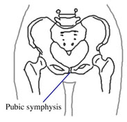
The pubic symphysis is the joint located between the two bones of the pelvis. In mammals, the pelvis consists of two large bones that support the spinal column and the hind limbs. These two bones join together at the front at the pubic symphysis, or the interpubic joint.
Female mammals give birth through the pelvis. In some species, the pubic symphysis relaxes during pregnancy under the influence of the hormone relaxin. This loosening of the pubic symphysis allows the separation of the pelvic bones, and hence widens the birth canal and provides an easier passage of the infant during birth.
Such relaxation of the pubic symphysis during pregnancy occurs in humans, guinea pigs and bats (Thoms 1936, O'Connor et al. 1966, Steinetz et al. 1983, Talmage 1947). For example, just before birth in guinea pigs, the pubic symphysis separates to a gap of 15 mm (Sutherland and Festing 1987).
In some species, the pubic symphysis is gradually replaced by bone with age. This means that after a certain age the pubic symphysis can no longer relax during birth. In guinea pigs, the pubic symphysis fuses in males and in females not bred before 6 months of age (Smallwood 1992). In female guinea pigs bred for the first time after 7 months, the pubic symphysis cannot separate, leading to abnormal, difficult, or life-threatening labor (Woerpel and Rosskopf 1988).
What's the story of the rat's pubic symphysis? Does it relax during birth? Does it turn to bone with age?
The rat's pubic symphysis
From birth through the second month, the rat's pubic bones rapidly ossify toward the pubic symphysis. By eight weeks of age, the pelvis and pubic symphysis have reached the adult condition: the pubic symphysis is a narrow band of cartilage 1.5 to 2 mm wide between ossified pelvic bones (Ruth 1935).
Adulthood: from sexual maturity to old age
The pubic symphysis changes very little during adulthood. There is almost no difference between the pubic symphysis at 6 months, 12 months, and 18 months, with only a slight narrowing as the rat grows older (Ruth 1935).
In pregnant rats, the hormone relaxin is present in the blood (Sherwood 1994), where it promotes a number of physiological changes such as the growth of the vagina and cervix during pregnancy (Burger and Sherwood 1995), and facilitiates delivery (Zhao and Sherwood 2004).
However, the female rat's pubic symphysis appears to relax only slightly (Crelin and Brightman 1957), or not at all (Ortega et al 2001) during birth. The pubic symphysis is the same in unmated, birthing, and post-partum rats (Ortega et al. 2001, but see Samuel et al. 1996, 1998).
These findings imply that the female rat's unaltered pelvis is large enough to permit the passage of infant rats during birth.
In aging male and female rats, the cartilage of the pubic symphysis degenerates and is gradually replaced by bone. By age 30 months the process is well underway, though the joint has not yet entirely fused by this time (Ruth 1935).
Conclusion
The rat's pubic symphysis does not appear to relax very much, if at all, during birth. The female rat's unaltered, adult pelvis appears wide enough to permit the passage of infant rats during birth. In elderly rats the cartilage of the joint gradually degenerates and is replaced by bone, but this occurs after menopause (15-18 months) in female rats.