- Introduction
- Are there disorders in laboratory rats similar to the Australian blue mutation?
- Can humans have this disorder too?
- What about other species?
- What do these syndromes have in common?
- Does the Australian blue mutation cause a disorder in lysosome-related organelles?
- Appendix: Chediak-Higashi and Hermansky-Pudlak syndrome in other species
Introduction
|
|
|
Spritely, an Australian blue hooded rat. Spritely died at a young age of a lymphosarcoma-like condition. His story was the inspiration for this page. Photograph reproduced with permission of the Dapper Rat . |
Possible human analogues and similar conditions were discussed, such as von Willebrand disease, hemophilia, and warfarin resistance. However, none of these suggestions had much explanatory power. The first two of these suggestions, von Willebrand disease and hemophilia, are blood-clotting disorders. They do not explain the other traits of Australian blue rats such as their depressed immune function and why these symptoms were associated with the blue fur color.
The third suggestion, warfarin resistance, is a heritable trait that renders blood resistant to the blood thinner warfarin (commonly used in rat poisons). There is also evidence that the warfarin resistance mutation may impair a blood clotting factor (Rost et al. 2004). However, as with von Willebrand disease and hemophilia, warfarin resistance would not explain the observed immunodeficiency or hypopigementation of Australian blue rats.
Are there any possibilities that might explain the whole suite of symptoms displayed by Australian blue rats, and not just their bleeding disorder?
Yes. Disorders in lysosome-related organelles give rise to individuals with dilute pigmentation, depressed immune function, and prolonged bleeding. This rare type of disorder has been found in numerous species including rats, mice, mink, cattle, cats, humans, and killer whales. It affects the development and function of certain "lysosome-related" organelles inside cells, particularly in cells involved in the immune response, pigment production, and blood clotting.
A disorder in the development and function of these lysosome-related organelles can be caused by different mutations in different genes. The mutations give rise to slightly different syndromes, such as Chediak-Higashi syndrome and Hermansky-Pudlak syndrome. I hypothesize that the Australian blue mutation may be a disorder of lysosome-related organelles that gave rise to a disorder similar to Chediak-Higashi syndrome.
Are there disorders in laboratory rats similar to the Australian blue mutation?
In other words, are there disorders that cause reduced pigmentation, prolonged bleeding, and impaired immune function in laboratory rats?
Yes.
In 1989, Nishimura et al. described a new mutation that appeared in an inbred colony of rats maintained at Hamamatsu University School of Medicine, a colony that originated at the Australian National Institute of Genetics. These rats displayed dilute pigmentation and prolonged bleeding times. After the amputation of the tail tip, normal rats bled for about 5-6 minutes before clotting, while the mutant rats did not stop bleeding even after 15 minutes. These rats also showed a great reduction in the natural killer function of their immune systems, though they did not appear particularly susceptible to infections in the laboratory.
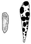
|
|
|
Giant granules in mast cells of beige rats (right) and normal rats (left). Drawn from Nishimura et al. 1989. |
The authors named the autosomal recessive mutation beige, after the diluted coat color of the rats and a similar mutation in mice, and suggested that this mutation was similar to Chediak-Higashi syndrome found in humans and other species.
Further studies
Further examination of these beige rats revealed that they had giant granules in a wide variety of tissues, including the liver, kidney, lung, bone marrow, peripheral blood, stomach, intestine, skin, and thyroid (Ozaki et al. 1994).
Beige rats also frequently developed spontaneous skin lesions, such as crust formations, itchiness, excoriation, hair loss, erosion and ulceration (Ozaki et al. 1997, 2005).
The bleeding disorder of beige rats was further characterized as a storage pool deficiency. Beige rats had a normal number of platelets, but these platelets were less able to aggregate to form a clot. Platelets of beige rats had fewer dense granules and lower levels of platelet ADP and serotonin (Ozaki et al. 1998).
In the laboratory, beige rats survived into old age, but when tested experimentally they were more susceptible to parasitic infection than normal rats (Nishimura et al. 1990).
The beige mutation was eventually mapped to chromosome 17 to the Lyst locus (Nishikawa and Nishimura 2000).
A second spontaneous beige mutant
Another mutation causing coat color dilution, prolonged bleeding times, and enlarged granules appeared independently in another strain of rats in 2003 (Mori et al. 2003), which turned out to be a second, spontaneous mutation in the same beige gene (Masui et al. 2004).
Can humans have this disorder too?
Yes.
Chediak-Higashi syndrome
Individuals with CHS are characterized by reduced pigmentation (silvery blond hair, pale blue eyes), prolonged bleeding, severe immune deficiency, and progressive neurological dysfunction. Individuals also have recurrent sinus and lung infections, and recurrent skin infections (superficial pyoderma to deep subcutaneous abscesses and ulcers). They may also show fever unrelated to an infection, and gum disease (eMedicine, OMIM, Dell'Angelica et al. 2000, Shiflett et al. 2002, Ward et al.2002, Windhorst and Padgett 1973).
Mortality is high. Death usually occurs before age ten, though some individuals have survived into their teens and twenties. Death is usually from an infection, bleeding, or development of an accelerated lymphoma-like phase.
Eighty percent of patients undergo the accelerated lymphoma-like phase. This phase is a non-malignant infiltration of multiple organs resembling lymphoma (lymphosarcoma), a cancer of the lymphatic system. The accelerated phase is triggered by viruses and leads to overwhelming infections of the skin, lungs, and respiratory system followed by death. CHS is inherited as an autosomal recessive (eMedicine, OMIM, Dell'Angelica et al. 2000).
The beige mutation in rats is thought to be an analogue of Chediak-Higashi syndrome in humans.
Hermansky-Pudlak syndrome
Individuals with HPS are characterized by a partial lack of pigmentation (blond hair and pale skin, though some may have brown hair and brown eyes), prolonged bleeding, and problems with cellular waste disposal: specifically, lysosomal ceroid storage (causes a build-up of a wax-like substance in cells). Lysosomal ceroid storage problems may cause pulmonary fibrosis (scarring of the lungs), inflammatory bowel disease (includes diarrhea, weight loss etc.), and kidney disease. Most affected individuals are legally blind due to lack of pigmentation in the eyes (eMedicine, OMIM, Dell'Angelica et al. 2000).
About 70% of affected individuals die from complications from HPS. Pulmonary fibrosis causes death in half of the individuals with HPS, usually in their 30s. Bleeding leads to the death of 10% of affected individuals. Other causes of death include intestinal, liver, and kidney failure (eMedicine).
Additional related syndromes are:
- Griscelli syndrome: characterized by dilution of skin color and silvery hair, severe immunodeficiency, and neurologic impairment. Average age at death is 5 years (eMedicine)
- Elejade syndrome: characterized by silver-leaden hair and neurologic impairment (seizures, hypotonia, developmental delay). Immune function is normal. Death is caused by neurologic problems and usually occurs during chilhood (eMedicine)
What about other species?
Chediak-Higashi and related syndromes have been found in a wide variety of species in addition to humans and rats, including mice, mink, cattle, foxes, cats, and killer whales (see below for more detail).
The syndromes are not identical between the different species, but tend to show reduced pigmentation of the hair and skin, prolonged bleeding, and in some species an increased susceptibility to infection.
What do these syndromes have in common?
Chediak-Higashi and Hermansky-Pudlak syndromes are examples of disorders in lysosome-related organelles.
Cells have tiny organs inside them that carry out a variety of functions. These little organs are called organelles. On a smaller scale, organelles are to cells what organs are to the body: organelles are the organs of a cell.
These organelles can be organized into families: a group of organelles that share a common lineage and features. One such family of organelles is the lysosome-related organelles.
Lysosome-related organelles are a family of organelles that are bound to inner membranes of the cell, have an acidic internal environment, and contain acid-dependent enzymes. They are found in many different cell types and have a number of functions. Specifically, lysosome-related organelles are involved in waste processing in the cell, pigment production, blood clotting, and the immune response (Table 1) (Dell'Angelica et al. 2000).
Table 1. Lysosome-related organelles and their functions (adapted from Dell'Angelica et al. 2000)
Lysosome-related organelle
Involved in:
Function
Lysosomes
Waste removal
Remove and digest waste from the cell. Found in all cells.
Melanosomes
Pigment production
Synthesize and store melanin pigments in pigment cells. Carry the pigment to the edge of the cell for deposit in the growing hair.
Platelet-dense granules
Blood clotting
Play a critical role in blood clotting. Secreted from blood platelets.
Lytic granules
Immune response
Secrete macromolecules which enable cytotoxic T lymphocytes (type of white blood cell) to destroy virus-infected or tumor cells.
MHC class II compartments (MIICs)
Immune response
Location of the binding of antigens to the MHC complex in antigen-presenting cells of the immune system.
Neutrophil azurophil granules
Immune response
Contain bacteria-killing enzymes. Found in neutrophils (type of white blood cell) which play a central role in defense against invading bacteria.
Basophil granules
Immune response
Contain inflammation effectors (e.g. histamin). Located in basophils, which are major cellular mediators of inflammation associated with allergic diseases.
Many genes are involved in the development and function of this family of lysosome-related organelles. A mutation in one of these genes will therefore have a wide variety of consequences, such as disorders of pigment production, blood clotting, and immune function.
|
Examples of genes involved in disorders of lysosome-related organelles Chediak-Higashi is caused by a defective lysosomal trafficking regulator termed CHS1 (formerly called LYST), located on chromosome 1q42-43. CHS1 is involved in fusion/fission events that determine the size of lysosomes and lysosome-related organelles. CH1 is also involved in protein transport to and from lysosome organelles in cells. Affected individuals have giant lysosome-related organelles (lysosomes of white blood cells, azurophilic granules, melanosomes in pigment cells) (OMIM, Dell'Angelica et al. 2000). The same gene may be involved in Chediak-Higashi in different species (Perou and Kaplan 1993, Shiflett et al. 2002). Hermansky-Pudlak: Caused by abnormal melanosomes, absence of platelet dense granules, and abnormal lysosome function. There are several different types of Hermansky-Pudlak syndrome, associated with different gene loci in humans: HPS1 plays a role in biogenesis of lysosome-related organelles, and ADTB3A mediates trafficking of membrane proteins to lysosome-related organelles. There are 16 mutations affecting at least six different proteins associated with HPS in mice (e.g. pale ear, pearl, mocha, pallid, gunmetal etc.) (Dell'Angelica et al. 2000, Li et al. 2004). |
Is the Australian blue mutation a disorder of lysosome-related organelles?
Nobody knows the answer to this question for sure, and we will not know until further studies have been done.
However, the constellation of symptoms displayed by Australian blue rats -- hypopigmentation, prolonged bleeding, and immune deficiency (respiratory and skin infections) -- are strongly suggestive of a disorder in lysosome-related organelles that affects melanosomes, platelet dense granules, and related organelles of cells involved in immune function.
The Australian blue mutation might, or might not, involve the same gene as the beige mutation found in laboratory rats, but Australian blue may well affect the same family of lysosome-related organelles, thus leading to a constellation of symptoms similar to Chediak-Higashi and Hermansky-Pudlak syndromes in humans and other species.
Testing this hypothesis
A simple test of this hypothesis could start with a non-invasive blood and hair sample from Australian blue rats and normal rats, followed by proper staining and examination under a light microscope. Hair could be examined for sparse, enlarged, clumped melanin granules in Australian blue rats as compared to the numerous, small, scattered melanin granules of normal rats. Suitably stained white blood cells such as neutrophils could be examined for enlarged lysosome-related organelles, when compared to those of normal rats.
For example, the protocol followed in Kramer et al. 1977 for the study of hair and blood samples in Persian cats with Chediak-Higashi syndrome could be adapted for use in rats. See also Prieur and Collier (1981) for a morphologic comparison of the melanin granules in the hairs of cats with different coat-color dilutions, including Chediak-Higashi.
Additional studies mentioned in this article contain more invasive methods for diagnosing Chediak-Higashi syndrome.
Testing the hypothesis that the Australian blue mutation in rats causes a disorder in lysosome-related organelles similar to Chediak-Higashi syndrome might be suitable for a small project such as an undergraduate honors thesis. If anyone conducts this study, please acknowledge me and contact me with your results.
Appendix: Chediak-Higashi and Hermansky-Pudlak syndrome in other species
Mice
Chediak-Higashi syndrome in mice: Several mutations have been identified in mice that are homologous to Chediak-Higashi syndrome in humans. The first mutant mouse was induced fifty years ago: it was on an agouti and black background and showed reduced pigmentation of the fur and skin (Kelley 1957, Davisson and Lewis 1990). These affected mice were beige colored, so their mutation was named beige (Withan and Lane 1991).
A second spontaneous mutation arose later on a non-agouti and black background. These mice have charcoal-grey fur and are the most commonly used "beige" mice in the laboratory today (Witham and Lane 1991).
Affected mice have reduced pigmentation in their hair and skin, and eye color ranges from ruby to almost black. The actual fur color depends on the genetic background (Sundberg 1992).
The melanin granules of beige mice are large and clumped but few in number, leading to a diluted coloration of the hair and skin. A wide variety of cells have giant granules, including many types of white blood cells, and cells of various organs such as the kidney and lungs (Lutzner et al. 1967, Sundberg 1992). As they age, affected mice display a progressive neurological disorder (Murphy and Roths 1978).
Beige mice are immune deficient, with deficiencies in their ability to kill bacteria, in the cytotoxic T-cell antibody response to tumors, and in the function of their natural killer cells. They are therefore more susceptible to infectious diseases. Heterozygotes also exhibit prolonged bleeding in the form of a storage pool deficiency. Beige mice weigh less and die younger than normal mice (Sundberg 1992).
Hermansky-Pudlak syndrome in mice:
Hermansky-Pudlak syndrome is characterized by hypopigmentation (e.g. blue fur, ruby eyes), poor vision, prolonged bleeding, and early death from lung disease. These traits are caused by abnormal pigment cells, abnormal dense granules in blood platelets, and abnormal waste disposal from cells. The shade of fur color and the degree of dilution depend on other background coat color genes (Li et al. 2004).
There are multiple forms of Hermanskly-Pudlak syndrome in mice. Each is caused by a different mutation. All of them affect the development of a lysosome-related organelles.
- Photos of Hermansky-Pudlak,
Chediak-Higashi, and control mice (Li et al. 2004)
- Chediak-Higashi mouse is Lyst(bg-J) (third row, fourth column)
- Control mice are C57BL/6J (row 1 counting from the top, column 1), C3H/HeJ (row 4, column 1), and DBA/2J (row 5, column 3)
- The rest of the mice are various genotypes of Hermansky-Pudlak syndrome in mice
Mink
Chediak-Higashi syndrome was found in mink with the pale, blue-grey, gunmetal coat color called "Aleutian." Pelts from these animals are called "Blue Iris" (e.g. blue iris mink hat and coat and earmuffs). Blue iris pelts are more valuable than normal ones, so mink with this trait were preferentially bred (Padgett et al.1964).
Aleutian mink have abnormal granulations in their leukocytes (white blood cells) and many cytoplasmic inclusions (Padgett et al. 1964, Lutzner et al. 1965). The disorder is inherited as a simple recessive: homozygous individuals display the disorder while heterozygous carriers show no symptoms (Padgett et al. 1964).
The leucocyte disorder was found in minks that were homozygous for Aleutian. These minks were not all pale, gunmetal, Aleutian blue: some were saphire, triple pearl, red-eye hedlund white, lavender, blue iris, hope, and winterblue. However, all of these mink phenotypes are homozygous for Aleutian and all are pale in color. Their different shades of coat color depended on the presence other coat color genes in addition to Aleutian (Padgett et al.1964).
Aleutian mink are weaker than normal mink: they have smaller litters of weaker kits (Helgebostad 1963) and are prone to skin abscesses (American Fur Breeder, 1961, p. 111).
Cattle
Chediak-Higashi syndrome has been identified in several different breeds of cattle:
Hereford cattle: Affected cattle are white with hypopigmented grey eyes and a faint fawn color where the normal red Hereford color pattern occurs. These cattle have abnormal granulations in their leucocytes (white blood cells). Cattle showed an increased susceptibility to disease. None of the affected individuals survived beyond four years of age (Padgett et al. 1964).
Japanese black cattle: Affected cattle have partial albinism and show a prolonged bleeding time (Shiraishi et al. 2002). The gene responsible is found on bovine chromosome 28, at a location homologous to Chediak-Higashi in humans (Kunieda et al.2000).
Brangus cattle: Chediak-Higashi was reported in three Brangus calves (Ayers et al. 1988). The affected calves had a hypopigmented, pale grey coat instead of the normal black color for that breed, and their eyes were grey instead of the normal dark brown to black. One of the calves displayed an abscess on the left side of the jaw and an ulcer at the base of the tongue. The second calf bled for an abnormally long time (30 hours) in response to a minor scrape, and at 18 months developed a massive bruise probably resulting from a kick. Certain types of the calves' white blood cells had abnormal, enlarged granules and the hairs showed abnormal pigment distribution.
Foxes
Nes et al. (1983) describe a Chediak-Higashi-like syndrome in Arctic blue foxes (Alopex lagopus). Affected foxes had greatly prolonged bleeding times in response to minor injury. These foxes had normal numbers of platelets, but the aggregation of these platelets was impaired (Sjaastad et al. 1990).
Killer whales
Chediak-Higashi was diagnosed in a young, wild killer whale in the early 1970s. The whale was brought into captivity, where it was named "Chimo" (photos). It was found to have partial albinism: photographs indicate that its skin was pale grey where normal killer whales are black. It had increased susceptibility to bacterial skin infections, enlarged granules in its white blood cells (neutrophils), and enlarged pigment granules in its pigment cells. It lived for two years in captivity then died of a streptococcal infection which developed into pneumonia (Haley 1973, Taylor and Farrell, 1973).
Cats
Kramer et al. (1975) report on a Chediak-Higashi-like disorder in a line of Persian cats. Affected cats had "blue smoke" colored fur and yellow eyes, abnormally large granules in the neutrophils of their blood and bone marrow, and enlarged melanin granules in their hair and skin. Their hair shafts had large, long clumps of melanin instead of numerous small, scattered, dark brown granules of melanin found in normal hair. This skin had few but abnormally large melanin granules. Many of the cats also developed bilateral nuclear cataracts by three months of age (Kramer et al. 1977).
The affected cats bled more profusely and for a longer period of time after a blood draw than normal cats. Affected cats also developed bruises after blood collection, while normal cats did not. The cats showed no increase in susceptibility to disease, but the number of cats examined was very small, so one cannot conclude that Chediak-Higashi cats are as resistant to infection as normal ones (Kramer et al. 1977).
It is possible diagnose Chediak-Higashi syndrome prenatally in cats, by examining fetuses for enlarged granules in the neutrophils (a type of white blood cell) in fetal blood (Kahraman and Prieur 1989), and enlarged lysosomes in chorionic tissue (Kahraman and Prieur 1991) and amniotic fluid (Kahraman and Prieur 1990).
Comparisons
See Windhorst and Padgett (1973) for a table on comparative aspects of Chediak-Higashi syndrome in humans, mink, cattle and mice.